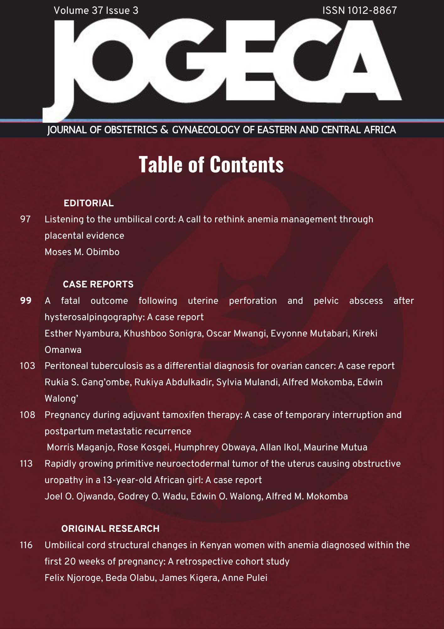
Listening to the umbilical cord: A call to rethink anemia management through placental evidence
DOI:
https://doi.org/10.59692/jf0b3776Abstract
There is untold story in every placenta, that within lies a biological record of pregnancy including signatures of maternal health, fetal adaptation, and the often-unseen consequences that arise when physiological demands outpace supply. Njoroge and colleagues in this issue present compelling evidence that early pregnancy anemia has potential to leave measurable, lasting imprints on the umbilical cord and I dare say the future human being. These findings challenge us to reconsider both the timing and the approach to managing anemia associated with pregnancy in clinical practice (1).
Gestational anemia affects over half of pregnant women in Kenya and many other countries in Africa (2). WHO classifies anaemia as a severe public health concern when its prevalence exceeds 40% (3). Yet despite decades of iron supplementation programs, antenatal screening programs, and public health campaigns, the needle has barely moved. Perhaps the reason lies not in the interventions themselves, but in their timing.
The study by Njoroge et al. examined umbilical cords from 36 women, comparing those diagnosed with anemia before 20 weeks gestation to those with normal hemoglobin levels (1). They reported significant differences in cord diameter, Wharton’s jelly volume, and umbilical vein wall thickness between the two groups. These are not subtle histological findings; they may as well represent fundamental alterations in the very conduit that sustains fetal life.
The umbilical vein obtained from women with anemia showed intima-medial thickening. This is normally a compensatory response to altered blood flow dynamics (1). More concerning is the disruption and reduplication of the internal elastic lamina, particularly pronounced at the fetal end of the cord. These structural alterations closely parallel those observed in systemic vascular disease, such as atherosclerosis and diabetic vasculopathy where sustained physiological stress drives adaptive remodeling that, over time, compromises vascular integrity and function.
Consider the implications. The umbilical vein delivers oxygen-rich, nutrient-laden blood from the placenta to the developing fetus. When its walls thicken and stiffen, compliance decreases. The vessel’s capacity to regulate and accommodate fluctuations in blood flow becomes limited. Coupled with reduced Wharton’s jelly volume, which normally cushions and protects umbilical vessels (4-5), and we have an anatomical explanation for why early anemia translates into adverse pregnancy outcomes (6-7).
The finding that these changes are more pronounced toward the fetal end of the cord is particularly noteworthy. The changes may be associated with a higher expression of vascular endothelial growth factor (VEGF) in this region, a compensatory response to hypoxia, (1). The fetus, most dependent on optimal umbilical blood flow, bears the greatest structural burden of maternal anemia.
References
1. Njoroge F, Olabu BO, Kigera JWM, Pulei AN. Umbilical cord structural changes in Kenyan women with anemia diagnosed within the first 20 weeks of pregnancy: A retrospective cohort study. J Obstet Gynaecol East Cent Afr. 2025;37(3):116-124. doi:10.59692/jogeca.v37i3.503
2. World Health Organization. Anaemia [Internet]. Geneva: WHO; 2023 [cited 2026 Mar 16]. Available from: https://www.who.int/news-room/fact-sheets/detail/anaemia
3. Tirore LL, Areba AS, Tamrat H, Habte A, Abame DE. Determinants of severity levels of anemia among pregnant women in Sub-Saharan Africa: multilevel analysis. Front Glob Womens Health. 2024;5:1367426. doi:10.3389/fgwh.2024.1367426
4. Heil JR, Bordoni B. Embryology, Umbilical Cord. In: StatPearls [Internet]. Treasure Island (FL): StatPearls Publishing; 2023 [cited 2026 Mar 16]. Available from: https://www.ncbi.nlm.nih.gov/books/NBK557490/
5. Debebe SK, Cahill LS, Kingdom JC, Whitehead CL, Chandran AR, Parks WT, et al. Wharton's jelly area and its association with placental morphometry and pathology. Placenta. 2020;94:34-38. doi:10.1016/j.placenta.2020.03.008
6. Chen Y, Zhong T, Song X, Zhang S, Sun M, Liu X, et al. Maternal anaemia during early pregnancy and the risk of neonatal outcomes: a prospective cohort study in Central China. BMJ Paediatr Open. 2024;8(1):e001931. doi:10.1136/bmjpo-2023-001931
7. Figueiredo ACMG, Gomes-Filho IS, Silva RB, Pereira BLS, Mata FAFD, Lyrio AO, et al. Maternal anemia and low birth weight: A systematic review and meta-analysis. Nutrients. 2018;10(5):601. doi:10.3390/nu10050601
8. Khong TY, Mooney EE, Ariel I, Balmus NCM, Boyd TK, Brundler MA, et al. Sampling and definitions of placental lesions: Amsterdam Placental Workshop Group Consensus Statement. Arch Pathol Lab Med. 2016;140(7):698-713. doi:10.5858/arpa.2015-0225-CC
9. Redline RW. Four major patterns of placental injury: a stepwise guide for understanding and implementing the 2016 Amsterdam consensus. Mod Pathol. 2021;34(6):1074-1092. doi:10.1038/s41379-021-00747-4
10. Correa-Agudelo E, Kim HY, Musuka GN, Mukandavire Z, Miller FD, Tanser F, et al. The epidemiological landscape of anemia in women of reproductive age in sub-Saharan Africa. Sci Rep. 2021;11:11955. doi:10.1038/s41598-021-91198-z
Downloads
Published
Issue
Section
License
Copyright (c) 2025 Moses Obimbo

This work is licensed under a Creative Commons Attribution 4.0 International License.